|
|
|
|
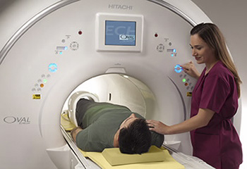
The Widest Wide-Bore in the Industry
Designed around the shape of the body to accommodate the broadest patient spectrum, Echelon Oval embodies our innovative spirit and unwavering commitment to patient comfort without compromising image quality.
Echelon Oval brings together all the key attributes of a leading 1.5T MR system – patient accessibility, workflow, and clinical capabilities all covered by Hitachi’s industry leading customer support.
● The widest patient opening (74cm) of any 1.5T MR system


Software Advancing Patient Care, Capabilities Advancing Your PracticeEvolution 6 is a robust software upgrade, offering over 30 new features and improvements to increase the performance of your Echelon Oval 1.5T platform. These new features allow your system the ability to remain at the forefront of technology. Operability and image quality improvements are included to help with overall workflow and system throughput. Evolution 6 introduces new options that add comfort, diagnostic breadth and insight to your portfolio of MR applications including Sound Reduction, Quantitative Mapping, Dynamic Imaging and more. HiMARAdvanced Metal Reduction with Hitachi’s isotropic voxel high resolution acquisition (isoFSE) SoftSound Brings quiet scanning for head to toe scanning FatSep TRAQ Multi-phase dynamic imaging in a single breath hold |


|
The WIT architecture combines novel technology and software enhancements that deliver workflow efficiencies at each stage of the exam. RF System A 16 or 32 channel RF system with integrated Head and Body/Spine coils can be used individually or in combination with anterior coils for comprehensive neuro and body imaging. Light-weight coil designs, multiple coil connection ports, and auto element selection all contribute to simplify patient positioning and extended coverage in head-first and feet-first imaging for all scans. WIT Monitor The WIT monitor on the face of the gantry provides patient information and assures gating integrity without repeated trips to the control room. The technologist can view basic patient information, patient orientation, coil set-up and confirm gating functions are working properly all right at the gantry, reducing the technologist tasks prior to starting the scan. Mobile Table A key contributor to smooth workflow is Echelon Oval’s WIT Mobile Table - the widest available tabletop in 1.5T. Patients are prepped and comfortably positioned in the exam room as the technologist confirms patient and scan information. Translation…more completed scans done faster. ● 63 cm wide – widest available ● 550 lb. weight capacity ● 20” minimum elevation ● Elevation adjustable when detached Digital Drive Digital Drive DX provides analog to digital conversion of the RF signal right at the gantry with fiber optic transmission to the equipment room to reduce signal loss and noise pick-up. With the digital conversion in the scan room, the MR signals are isolated from the external noise sources, resulting in the highest possible signal to noise ratio. Origin Echelon Oval’s Vertex II computer and Origin MR operating software deliver seamless workflow from patient registration through post processing. The Clinical Study Library, Graphical User Interface (GUI), Intelligent Parameter Guidance, and Real-time Image Quality Calculator simplify acquisition planning for even the most complex examinations. Features: ● Simultaneous scan, reconstruction, and multi-tasked image processing ● HIS/RIS Interoperability ● DICOM and IHE (SWF-PIR) compliant ● Automatic protocol selection ● Intelligent parameter guidance ● Real-time image quality calculator |


|
|
Echelon Oval delivers the full spectrum of clinical capabilities. Our leadership in imaging techniques such as fat suppression, non-contrast MRA, and motion compensation combined with Hitachi exclusive capabilities ensure diagnostic confidence for even the most challenging patients. Neurological Capabilities That Matter Echelon Oval provides the essential acquisition and post processing tools for high-quality imaging of the brain and spine. ● High resolution, motion compensated visualization of anatomical structures for obtaining detailed images of the brain and nerve tissues with All Around RADAR suite including RAPID-RADAR for the diagnosis of lesions and disorders ● Fast and reproducible neurosvascular assessments with BSI, TOF and BeamSat TOF to evaluate blood vessels and blood flow ● Physiological and quantitative analysis provided by DWI, DTI, DKI and Perfusion imaging that deliver insights into characteristic differences of normal and diseased tissue Musculoskeletal Capabilities That Matter Taking advantage of the widest patient space for positioning closer to isocenter, Echelon Oval provides a flexible and robust platform for MSK imaging. ● opFSE provides high clarity and sharp images with superb edge detail for all Fast Spin Echo acquisitions ● Fast and effective metal artifact reduction techniques with primeFSE and FatSep ● Isotropic volume imaging with isoFSE to visualize challenging structures ● Multiple layer cartilage assessment with T2 Relax Map and microTE Body Capabilities That Matter Echelon Oval delivers enhancements in dynamic, diffusion weighted, and motion compensated imaging to aid diagnosis in a breadth of applications including liver, kidney, prostate and breast. ● Short breath-hold times with extended coverage for general body imaging with RAPID ● Extensive motion compensation techniques to reduce motion with the All Around RADAR suite of sequences ● TIGRE provides high temporal and spatial resolution with uniform fat suppression for dynamic imaging ● Whole Body DWI for lesion detection Vascular Capabilities That Matter Many factors including blood flow dynamics, bolus timing, and large FOV contribute to making vascular and cardiac imaging challenging even for the most experienced facilities. Echelon Oval provides easy to implement features that deliver consistent results head to toe. ● Simplify bolus timed MRA with FLUTE and TRAQ ● Non-contrast MRA solutions, VASC-ASL and VASC-FSE, reduce costs and risk ● Selective saturation MRA delivers controlled area of evaluation for brain and abdominal regions ● Functional morphilogical, perfusion and coronary artery imaging |


|

|
|
Magnet Echelon Oval’s 1.5T magnet features ultimate stability. Its design includes inherent shielding from magnetic fluctuations, high order active shimming technology (HOAST) applied per patient assuring exceptional magnetic field uniformity, and very low Helium boil off. ● 1.5 Tesla superconducting ● 50 x 50 x 50 cm FOV ● Guaranteed Homogeneity - 0.5ppm @40cm DSV (Vrms) ● Active magnetic shielding ● Helium cryogen ● Refill: Once every six years RF System Echelon Oval’s 16 or 32 channel RF receiver system manages multiple coil connection points on the table. The WIT RF coil system provides integrated coil arrays that can be used individually or in combination to give the operator maximum flexibility for positioning patients of all sizes. Virtually all of Echelon Oval’s array, surface, and volumetric coils are multiple element designs for high signal uniformity, high SNR, and compatibility with RAPID™ parallel imaging for maximum clinical flexibility and image quality. ● Two channel RF Transmit ● 40 kW peak power (2 x 20) ● Digital Receiver - 16 or 32 independent channels - A/D conversion on gantry with optical digital transmission - T/R body coil ● WIT Integrated Arrays and dedicated anatomy coils Gradient System Echelon Oval’s standard 34 mT/m, 150 T/m/s gradients deliver the capability for rapid acquisition of high-resolution images. The high peak strength and fast slew rate allow the user to select short TE times, small FOV, thin slices and high matrices. ● Peak amplitude: 34 mT/m ● Peak slew rate: 150 T/m/s ● Active shielding ● Water cooling ● Gradient noise reduction - Softsound™ sequences - Sound dampening Siting Echelon Oval’s design attributes make it accommodating to existing facilities and easily planned into new construction. As an acknowledged leader in imaging placements, Hitachi offers a wealth of site planning experience and a proven system for efficient siting, installation, and start-up. ● 5 gauss line - Axial 4.0 m (13' 1") - Radial 2.5 m (8' 2") ● Scan room size - 20’4” x 16’ 5” (Typical) - 19’7” x 13’2” (Minimum) |
Contact Prestige Medical Imaging Today To Address All Of Your X-Ray Imaging Questions